Previously, we wrote about how to “see” UV-C reflection or absorption. We’ve found that UV imaging can help us visualize how UV is scattered around a hospital room and, therefore, better understand which areas are being properly treated (and which may not be). While this is certainly useful, what we really need to know is the actual UV intensity on a given surface. In other words, how much UV light energy per square centimeter is hitting every surface in a room? This is the value that truly determines how much disinfection takes place. Scientists long ago correlated UV energy per area to germicidal performance, often expressed as log reduction.
UV intensity can be measured in a few different ways. Here we’ll discuss two: radiometric sensors and photochromic indicators.
Radiometric Sensors
Simply put, a radiometric sensor measures electromagnetic radiation. In our case, we are interested in the relatively narrow UV-C portion of the electromagnetic spectrum. Ideally, our sensor responds to UV-C wavelengths and ignores everything else. For example, we don’t care about the intensity of visible light, since those wavelengths are not germicidal. What is normally done is to add a filter that blocks out any wavelengths that we are not interested in.
Because germicidal irradiation is so prevalent in other industries, several companies make systems to measure UV-C intensity (usually referred to as “irradiance”). Normally, these consist of a sensor combined with a meter which converts the output from the sensor to a numerical value for irradiance in energy per area per time (e.g. Joules per square centimeter per second) . Below is the ILT1700 from International Light Technologies, which is the meter we use for most of our measurements.
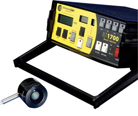
The sensor can be placed anywhere in the room and the results used to map out the UV distribution. It is especially important to understand how much UV is hitting areas that are not in direct line of sight of the device. In our experience, most surfaces that can see the device will get adequately disinfected in a few minutes. It is the shadowed areas that truly dictate how long a given device should run, but, unfortunately, most device manufacturers ignore this fact.
Photochromic indicators
A less exact, but simpler, method is to use a photochromic indicator. These typically are small adhesive labels containing special pigments that change color as they are exposed to UV. For example, they may change from an initial yellow color to a dark green. In the case of a product called Spectrify from Spectra254, the label is initially white and gradually turns to a dark pink color. The darker the color, the greater the UV exposure. Calibration standards have been created to show how the color corresponds to a particular level of exposure.

The advantage of this approach is that the labels are relatively inexpensive, so numerous labels can be placed around the room to measure UV in many locations simultaneously. The information received is only semi-quantitative, but, nonetheless, provides valuable insight into which areas of the room are being properly disinfected. Also, because of their size, these labels can be put into tight spaces in which a radiometric sensor may not fit.
The importance of UV measurements
UV device effectiveness data provided by manufacturers typically only includes surfaces that are directly illuminated by the device. This ignores the large number of surfaces that are illuminated only indirectly, or are completely shadowed. To understand the entire picture and to establish a more effective device cycle time, it is necessary to take additional measurements of these indirect surfaces.
However, we can’t ignore an important variable- the location of the device in the room. Changing the device location slightly may significantly change the UV dosage on some surfaces, as they move in or out of shadows. An important benefit of Lumacept is that the diffuse reflectance of the paint helps make the dependence on the device location less significant by providing much more indirect light and essentially “smoothing” out these differences.
To properly optimize the device location, many UV intensity measurements must be taken while systematically moving the device. This can be time intensive and tedious, which is why we developed LumaSim. Through computer simulations, we can take thousands of virtual measurements and easily study the effect of adjusting the device location until the best location is found. The combination of Lumacept coatings and an optimized device location can not only make your disinfection protocol more effective, it can also save significant time.
Have any technical or not-so-technical questions about UV? We’re happy to help. Contact us at info@lumacept.com.